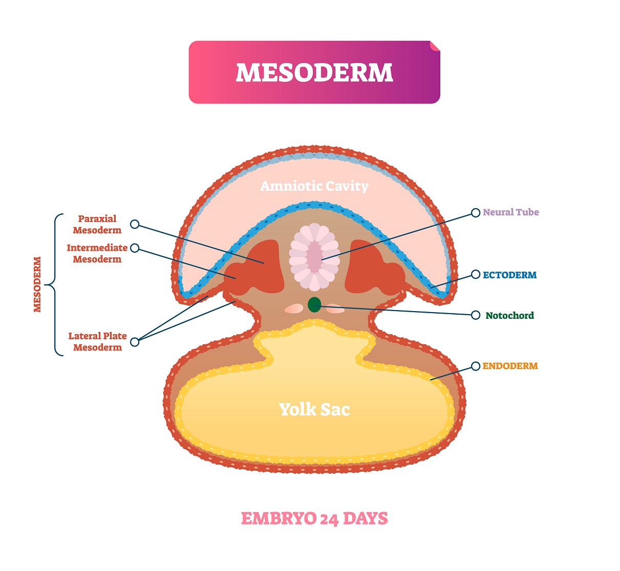
Part 1 of this series on the biology of pregnancy took us from conception through gastrulation, the process in which the cells of an early embryo arranged into three layers: The inner layer of cells, called the endoderm will form the inner linings of the baby’s gastrointestinal tract, the respiratory tract, and linings of various other other organ systems. The middle layer of cells, called the mesoderm, gives rise to muscle tissue and connective tissue all over the body. This means that mesoderm is vital to the development of bones and skeletal muscles and also to the heart and blood vessels, which we will discuss further, below. Finally, the outer layer of cells, called the ectoderm, is the source of the baby’s nervous system, plus other parts of the body, such as eyes, hair, the epidermis of skin, and the enamel of teeth.
Also early in development, during the third or fourth gestational week, another process, called neurulation, has begun. The initiation of neurulation occurred when a flat area of the embryo, called the neural plate, began to fold. By the fifth gestational week, the folding has caused the edges of the plate to approach one another in preparation to transform the plate into a tube, called the neural tube, that will close up as the edges of the folding plate meet up, along the midline of what will be the embryo’s back. In some areas along the embryo, the edges of the plate already have met up during the fifth week, but much of the developing neural tube is still open. Closure of the neural tube will occur gradually over the next couple of weeks and the neural tube will give rise to the central nervous system –the brain and the spinal cord. The process is vulnerable incomplete closure, however. Remaining gaps, called neural tube defects, can occur in the newborn, along the spine or skull, leaving parts of the spinal cord or brain unprotected. A major risk factor for the development of neural tube defects is a deficiency of the vitamin folic acid (folate). Although your obstetrician will instruct you to take supplemental folic acid pills (or pregnancy multivitamins containing adequate amounts of folic acid) to help prevent neural tube defects, if you are found to be pregnant, it is very unusual for a woman to know that she is pregnant during these early weeks of pregnancy. Consequently, you should be consuming adequate amounts of folic acid (by way of your diet and/or supplements), if you are having unprotected sex with the intention of becoming pregnant.
Even so, pregnancy is easily detectable by the 5th gestational week. This is a point when a woman who has normal menstrual cycles will know that she has missed her period. This might lead her to take a home pregnancy test, and/or to visit, either her primary care physician or an obstetrician/gynecologist (ob/gyn), either one of whom will perform an in-office pregnancy test, whether or not the woman has done a home pregnancy test. Usually, the woman’s care will continue with the ob/gyn from this point onward, but in some cases, especially in rural areas, a family physician with obstetric training may manage the case, until or unless there are complications.
Whether during the fifth week, or more commonly during the next few weeks, a positive pregnancy test leads to the next step, ultrasonography. Early on, your obstetrician will be looking for a gestational sac. By week five, there should be a yoke sac, and then comes something that aspiring parents are often excited to see and hear –a beating ‘heart’— although it’s not really a heart yet.
Over the next weeks, however, development of the cardiovascular system takes center stage. The heart begins as embryonic blood vessels fuse together, forming a muscular tube that is wider and carries more blood than any other embryonic vessels do. By the end of week five, the tube has twisted slightly and separated into two chambers –an atrium and ventricle—and it beats, propelling blood through the embryo. You’ll see the two-chambered heart as a pulsating feature in the center of embryo.
This emergence of a pumping tube that later gets divided into chambers is a re-play of the changes that took place in our ancestors over millions of years, but accelerated to occur in the span of a few months. At five weeks gestation, the two-chambered heart is almost exactly like the heart of a fish. But the architecture of the heart and the surrounding blood vessels will continue to change as the embryo grows. Genes are turning on and off in cells with precision accuracy, laying down the foundation for a kind of wall –called a septum—that will divide the single atrium into two. In the days to come, this will create a three-chambered heart (two atria and one ventricle), similar to what still exists in adult amphibians, like frogs.
Along with the shape of the heart, your doctor will also be looking closely at the rate at which the heart beats. When the heart pumping becomes visible around five weeks, the rate will be around 100-110 bpm. But over the next four weeks or so it will increase, up to 165-185. Then it will start decreasing from that point.