We’re now taking our journey through the biology of pregnancy from the second trimester to the third. The second trimester runs through the end of the 26th gestational week. This is a time when it’s particularly important for your doctor to screen for gestational diabetes, especially if you are obese, if you are older than 30, or if you have diabetes in your family. It’s also important for the doctor to check your legs for signs of deep venous thrombosis (DVT), a clot in a deep vein.
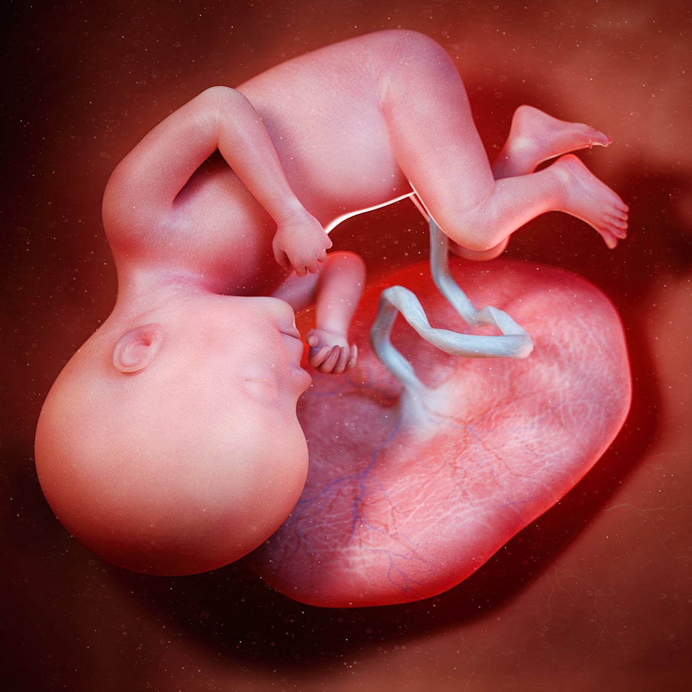
By this point, the basic body shape is set for the fetus, although it has a lot of weight to gain before it is ready for birth. Most internal organs are functioning as they will subsequent to birth, but with a few notable exceptions. One exception is the respiratory system. The fetus has no direct contact with the outside air, so no air is moving in and out of the lungs. Instead, the lungs are collapsed and bathing in amniotic fluid, which also serves to cushion the fetus within the womb. Pressure is very high in the pulmonary blood vessels and on the right side of the heart, but pressure on the left side of the heart is low. The right-left pressure difference causes most of the fetal blood to divert away from the lungs and into the aorta, the trunk-like artery that supplies the entire body. This is possible, because of shunts that carry blood from the right side of the heart into the aorta, the big artery that carries blood from the heart’s left ventricle to all of the arteries that nourish body tissues. Much of this blood comes from the left ventricle itself, after traveling from the right atrium, through an opening called the foramen ovale, through the left atrium, and through the mitral valve. Other blood, however, reaches the aorta by traveling from the right atrium, through the right ventricle, into the pulmonary artery and through another blood vessel called the ductus arteriosus. Only a very small amount of blood actually travels through the collapsed lungs themselves during fetal life, just enough blood to nourish the lungs.
Meanwhile, the situation of the blood pressure being high on the right side of the heart and low on the left side of the heart is exactly opposite of what happens after birth, when pressure becomes high on the left side and low on the right. But the fetal pressure situation is ideal during pregnancy, since the fetus gets oxygen from the mother’s lungs by way of the placenta. The oxygenated blood from the placenta enters the fetus through the umbilical artery that runs through the umbilical cord and continues into the fetal abdomen, but then there’s a fork in the road. One route carries most of the blood from the umbilical artery to the inferior vena cava, the large vein that delivers blood from the lower body of the fetus into right atrium. The other route carries blood from the umbilical artery through the blood vessels of the liver, but from the liver this blood also moves into the inferior vena cava, joining the rest of the blood on its way from the placenta, along with blood from the fetal legs.
Red blood cells are packages of hemoglobin –the protein that carries oxygen within red blood cells in blood. Fetal hemoglobin is slightly different from your hemoglobin. It holds oxygen just a little more tightly. Thus, when maternal blood and fetal blood get close together in the placenta, oxygen moves from the maternal blood into the fetal blood. At the same time, carbon dioxide moves in the other direction, from fetus to mother, and then out through the mother’s lungs. At this point in development, fetal red blood cells are produced mainly in the liver, and also in the spleen.
When the third trimester comes to a start with the opening of week 27, a lot is happening in the fetal brain. Invaginations, folds called convolutions, that characterize the outer layer of the cerebral cortex are getting more numerous and complex. The convolutions enables more surface area of nervous tissue to pack into the small cranial volume, but the brain also is removing what it doesn’t need. Nerve cells are dying to make room for others. Synapses –connections between neurons– continue to form at an accelerated rate, but many synapses are being snipped away. This neural pruning is a process that will continue throughout life. In childhood, and throughout adulthood, it’s an important part of learning. It’s a way of eliminating potentially distracting noise, like deleting unnecessary files, or clearing the cache on your computer, or mobile devise. Also, the developing left and right hemispheres of the cortex are now showing some anatomic asymmetry. Certain areas of the cortex are bigger in one hemisphere than in the other. As the child develops, the anatomic asymmetry increases, with consequences in function. The most notable consequence is handedness. Most people are right handed. The left side of the cerebral cortex controls motor function, if you are righty. If you are lefty, it’s the right side of the brain that controls your dominant hand. Speech and various other functions also depend on brain centers that are lateralized, meaning different between the left and right hemispheres.