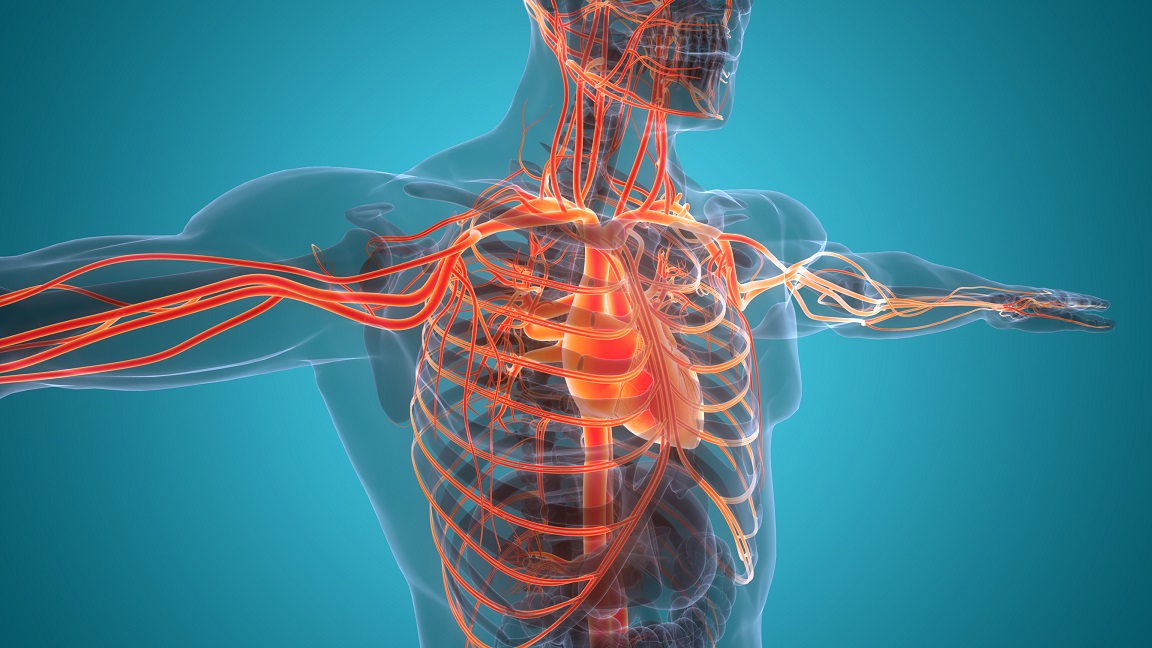
If you are pregnant, or are preparing to become pregnant, you probably know that your body is in for some changes. Changes in the shape and size of the body and its parts are obvious from the outside. We have known about these changes since before we were human, but you may be wondering about changes inside, changes in your physiology. There are many such physiological changes that develop as pregnancy progresses. Various body systems are affected and we will go through many of them in the course of a few blog posts. Today, let’s begin with the circulatory system, which consists of the heart, the blood vessels, the blood, the lymphatic vessels, and lymphatic fluid, also called lymph. Changes during pregnancy in the cardiovascular system (the heart and blood vessels) and in blood are numerous and many of the details are beyond the scope of a blog post, but let’s take a look at some of the main developments.
Changes in blood are rather dramatic. The volume of blood within your body increases by up to 50 percent by the end of pregnancy. This causes decreasing concentration in red blood cells (RBCs), causing a drop in the RBC count when your blood is tested, along with a drop in hemoglobin, which in many cases is accompanied by changes in various measurements in the blood that doctors use to determine if you are iron deficient. The body begins compensating for the dilution of RBCs by increasing RBC production. In some women, this acceleration in RBC production returns the RBC count and hemoglobin to normal, or close to normal by delivery, but often the RBC count remains low. This expansion of the blood volume compensates for your growing uterus, which requires a great deal of blood flow, for blood loss during delivery, and for expansion of blood vessels throughout the body to allow for blood to flow with decreased resistance. This is called a reduction in the systemic vascular resistance.
Accompanying the decrease in the systemic vascular resistance is an increase in the heart rate, meaning that your heart beats somewhat faster compared with your pre-pregnancy heart rate, typically up to 25 percent faster. Healthy, young women who are not pregnant typically have a heart rate in the range of 80 to 100 beats per minute. Your normal resting heart rate also may be a little bit below 80 beats per minute; if you are an endurance athlete, it could be a lot lower, even as low as 60 beats per minute. A heart rate above 100 is called tachycardia, but there are many different types of tachycardia. In the case of tachycardia that occurs during exercise, when you are excited, and during pregnancy, the tachycardia is physiological, meaning not part of a disease process, but merely a compensation for things happening in the body. The medical term for such a physiological acceleration of the heart rate is sinus tachycardia, which means that the rhythm of the heart beat and the pathways through which the electrical signals that control the heart rate and rhythm are normal. Normal means that the signal for the heart to beat begins within a concentration of specialized muscle cells in the heart’s right atrium called the sino-atrial (SA) node (a natural pacemaker), and that the impulse causes the two atria to contract, while also transmitting to another concentration of pacemaker cells called the atrio-ventricular (AV) node, which then, through a particular network of still more specialized muscle cells, tells the two ventricles to contract in a coordinated manner. While pregnant women normally experience only sinus tachycardia, occasionally there are some irregular heart rhythms that require women to be evaluated by a cardiologist, or an obstetrician who specializes in medical conditions during pregnancy.
Although the force of contraction of heart muscle (known as myocardial contractility) has not been shown to increase during pregnancy, the decrease in systemic vascular resistance enables blood to exit the left ventricle through the aortic valve to begin its journey through the body more easily than prior to pregnancy. Meanwhile, the increased blood volume leads to more blood returning from the body to the right side of the heart and passing through the lungs, into the left atrium, and into the left ventricle. These two factors lead to what doctors call an increase in cardiac output, which is defined as the volume of blood pumped out of the left ventricle each minute. Over the course of pregnancy, cardiac output gradually increases, peaking at 60 to 80 percent of normal by the time of labor and delivery and the hours following delivery.
Blood is also part of the circulatory system and it goes through some major changes as well. The tendency for blood to clot depends on three factors, known together as the Virchow triad, named for the famed, Prussian pathologist of the 19th century, Rudolf Virchow (1821-1902). One of these factors, damage to the inner lining of blood vessels, is a very big deal in the case of arterial blood clots involved in heart disease, strokes, and problems in the limbs and eyes in people with disease, but it is not so much an issue in most pregnant women. The other two factors of the triad, however, are very important. One is when blood slows down or stops moving within blood vessels, which tends to happen in the veins during pregnancy, because of your growing womb pushing on large, deep veins in the pelvis. The other consists of the cascade of various clotting proteins and other chemicals that enable the blood to form clots. During pregnancy, this clotting cascade shifts toward increased tendency of blood to clot. Particularly during the latter half of pregnancy, and for about six weeks after delivery, these changes put you at elevated risk for what’s called venous thromboembolism (VTE). This is a condition in which a clot forms in a deep vein, and in some cases may then cause a type of blood clot in the lung, called a pulmonary embolism. On the other hand, women who suffer from a fairly common disorder, called von Willebrand disease, in which they bleed too easily, tend to improve during pregnancy.