Newborns are often born with all kinds of markings on their skin, and many others develop different markings in the first few days or weeks of life. Are these all simply birthmarks? Or do they represent something truly worrisome? A few of the more common skin markings in newborns are presented here.
“Stork Bite”

One of the most common birthmarks is a nevus simplex, also known as a “stork bite” or “salmon patch.” This benign lesion is usually found in the center of the face: forehead, between the eyebrows, on either eyelid, and in the philtrum (the shallow groove between the nose and upper lip, as in the picture above), as well as the nape of the neck. It is usually a flat, pale pink to bright red lesion, and can look deeper red when the baby is crying or hot. Nevus simplex can be seen in 20-60% of newborns at birth, but fades between 1-3 years of age without any treatment. Sometimes, the nevus simplex on the nape of the neck can last much longer, even for life, but these are usually covered by hair.
Port Wine Stain

A nevus flammeus, also known as a port wine stain, are much less common, occurring in about 0.3% of newborns. Port wine stains are flat, red to deep red in color, and can be found anywhere, but are commonly seen on the face or head. They don’t resolve without treatment, and are usually just a cosmetic problem, but sometimes can signify problems with the blood vessels of the eye or brain. So if the port wine stain involves certain portions of the face (eyelid or maxillary areas), then the baby should be examined by an ophthalmologist and get some imaging of the head, such as an MRI. The other forms of port wine stain can be treated when the child is older with laser therapy.
Hemangioma
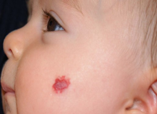
Often referred to as strawberry hemangiomas, these lesions are often bright red (like the fruit) and raised. They may appear anywhere on the body. Hemangiomas are common, occurring in about 10% of newborns, but are not always apparent at birth, often showing up a few weeks later. They usually grow in size for many months, but then slowly resolve over years. Sometimes the hemangiomas are deeper, and appear bluish or purple, and occasionally involve deeper structures, such as organs. But most hemangiomas are superficial, and require no treatment. Sometimes, larger hemangiomas or those that are located in a critical area are treated with an oral medication, propranolol, in order to shrink the hemangioma. Other, less common treatments include steroids, interferon, and laser treatment.
Nevus Sebaceous

Also known as a nevus sebaceous (or sebaceus) of Jadassohn, this lesion is usually a single, oval or linear-shaped, pink or yellow or orange, waxy, hairless, smooth, slightly raised lesion on the scalp, face or neck of a newborn. It occurs in 0.3% of neonates. During puberty, the nevus can change and become thicker and look more like a wart. A nevus sebaceous is usually an isolated finding, but at times can be a sign of more serious, systemic disease that includes neurologic, bone, and eye abnormalities. Previously, it was thought that these lesions led to skin cancer later in life, so were surgically removed at an early age. Currently, these lesions are usually observed for years before any surgical removal is considered.
Café-au-lait Spot

The name, the French word for “coffee with milk”, describes the color of these flat spots on light skin. Café-au-lait spots appear more like dark coffee color on dark skin. They are found anywhere on the body and are seen in 3-25% of newborns. They are usually isolated and benign, but can be associated with more serious syndromes, such as neurofibromatosis. If your baby has three or more of these spots, alert your pediatrician, who will monitor the baby for these other diseases.
Dermal Melanocytosis
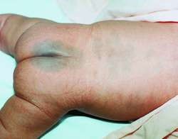
Also historically called a Mongolian spot, this flat, blue-grey lesion is commonly found in the lower back and buttocks area. It is seen in all races, but more frequently in Asian babies. It resolves spontaneously and does not need treatment.
Congenital Melanocytic Nevi

Congenital melanocytic nevi are also called moles, and are seen in about 1% of newborns. They are usually flat and vary in color at birth, but become thicker, often change in color, and can have hair growing from them as the child ages. They are usually small, isolated, and benign. However, very large nevi, those that will grow to >20 cm in size in the adult (>8 inches), have a higher risk of developing into melanoma, or skin cancer. A medical professional should watch these congenital nevi carefully. There is a rare syndrome of very large or multiple congenital melanocytic nevi that is associated with neurological symptoms, and these babies should have an MRI of their neurological systems.
Pigmentary Mosaicism
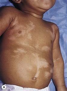
Pigmentary mosaicism presents as flat skin markings in a linear, whorled or segmental pattern. They are either darker or lighter than the normal skin, and occasionally both in the same child. This condition is usually isolated and benign, but can be associated with other abnormalities, including those of the heart, bones, muscles, and neurological system.
Speckled Lentiginous Nevus
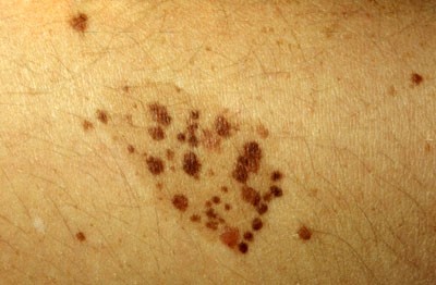
Also known as nevus spilus, the speckled lentiginous nevus is a solitary, flat, non-hairy, tan patch of skin with darker brown patches within. They can be found anywhere on the body, and occur in 1-2% of babies. They are usually benign; though sometimes develop into skin cancer as an adult.