Virtually every pregnant woman receives an ultrasound at some point during their pregnancy. And because it does not involve needles, it is considered a non-invasive prenatal test. Your doctor or sonographer uses a plastic transducer to transmit high-frequency sound waves through your uterus. These sound waves send signals back to a machine that converts them into images of your baby. The terms ultrasound and sonogram may be used interchangeably.
What does the ultrasound image look like?
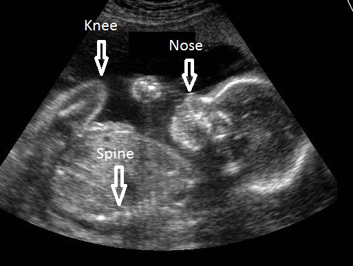
You will probably receive printed black and white photos from your ultrasound scan. On the photos, hard tissues such as bones reflect the most sound waves and appear white. Soft tissues such as cartilage and skin appear gray. Liquids such as amniotic fluid appear black because the sound waves go through liquids with no echoes.
Are ultrasounds safe?
Yes, as long as the machine is used following standard guidelines. Therefore, they have to be performed by a trained doctor or sonographer. Ultrasound does not use radiation like X-ray tests do.
What is an ultrasound scan used for?
Depending on your stage of pregnancy, ultrasounds can:
- Check that your baby has a heartbeat and whether it is normal
- Confirm whether you’re pregnant with one or more babies
- Check your baby’s heartbeat, muscle tone, movement and overall development
- Detect if the baby is implanted outside of your uterus (ectopic pregnancy), which would become a medical emergency
- Find out the cause of any bleeding you may be having
- Accurately date your pregnancy by measuring your baby from head to bottom (the calculation of your due date by ultrasound is the most accurate of all methods)
- Assess your baby’s risk of Down’s syndrome by measuring fluid at the back of your baby’s neck at 11 weeks plus two days to 14 weeks plus one day (this is called a “nuchal translucency” scan)
- Help with prenatal diagnostic tests, such as chorionic villus sampling or amniocentesis, by showing the position of the baby and placenta
- Examine your baby to see if all his organs are normal
- Diagnose most abnormalities, such as spina bifida and heart defects
- Assess the amount of amniotic fluid you have and find out where the placenta lies
- Measure your baby’s rate of growth over several scans
If you wish to know the gender of your baby, it can usually be determined by 20 weeks, but it’s not always possible to tell. Also, there is a chance that the ultrasound images can be misinterpreted.
At what stage in pregnancy are ultrasounds performed?
In the first trimester of pregnancy it can be done anytime. A gestational sac can be visualized on ultrasound as early as 4 ½ weeks after your last period; a heartbeat can be detected as early as 5 to 6 weeks (although it might not be detected that early in all cases).
Does an ultrasound hurt?
No. You may only feel mild discomfort from the transducer pressing on your abdomen, especially if you have a very full bladder. If you feel any pain, tell the sonographer.
Do I have to have an ultrasound?
Most women in the US have at least one scan during pregnancy, but you don’t have to have one. As I discussed above, scans give very useful information about your pregnancy, and most women find them reassuring.
How do I prepare for the ultrasound?
Sometimes, women are told to drink 4-6 glasses of water before the test, so your bladder is full, but not so full it causes pain. This helps the sound waves travel better through the skin and tissues. You may be asked to change from your own clothing into a medical gown.
What if the scan shows there’s a problem?
If the sonographer finds anything unusual during the scan, he or she will refer you to a doctor who may require further tests (such as chorionic villus sampling or amniocentesis).
Can a “normal” ultrasound miss a problem?
Yes. Finding an abnormality depends on many factors, including the age and position of the baby, as well as the size and type of abnormality.
What’s the difference between an ultrasound and Doppler fetal monitoring?
Doppler fetal monitoring will typically be performed during the last trimester if you suffer from gestational diabetes. A regular ultrasound uses sound waves to produce images; this one bounces high-frequency sound waves off circulating red blood cells to measure blood flow and blood pressure. The test will determine if your baby is getting enough blood.
What is a 3-D and 4-D ultrasound?
A 3-D ultrasound takes thousands of pictures at once. It makes a 3-dimensional image of your baby that is almost as clear as a photograph. Some health care providers use this kind of ultrasound to make sure your baby’s organs are growing and developing normally. You also may get a 3-D ultrasound to check for problems in the uterus. The 4-D ultrasound is like a 3-D ultrasound, but it also shows your baby’s movements in a video. Both types are safe for you and your baby as long as they are run by a health care provider.
I’ve seen that 3-D and 4-D ultrasounds are offered in a shopping center close to where I live. Is it safe to get one there?
According to the March of Dimes, the FDA, and other experts, the use of non-medical ultrasounds is discouraged because untrained personnel may provide inaccurate or harmful information.
Will my insurance pay for the ultrasound?
Insurance will pay for the ultrasound if it is deemed medically necessary. If you have an ultrasound that is not medically necessary (for example, to simply see the baby or find out the baby’s sex), your insurance company may not pay for the ultrasound.